|
Inhaltsübersicht | Nanomaschinen | Moleküle | Programme | Kurse | Fun | Links |
|
|
| > |
Hemoglobin
Exploring the Structure
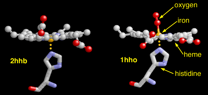
You can look at the binding of oxygen up close in two structures of human hemoglobin. PDB entry 2hhb shows hemoglobin with no oxygen bound. In this picture, the heme is seen edge-on with the iron atom colored in gold. You can see the key histidine reaching up on the bottom side to bind to the iron atom. In PDB entry 1hho, oxygen has bound to the iron, pulling it upwards. This in turn, pulls on the histidine below, which then shifts the location of the entire protein chain. These changes are transmitted throughout the protein, ultimately causing the big shift in shape that changes the binding strength of the neighboring sites.This picture was created with Rasmol. You can create similar pictures by clicking on the accession codes and choosing one of the options under View Structure. Note that the PDB entry 1hho only contains two of the four chains in the hemoglobin structure. To get the full tetramer, click on the accession code, and then go to Other Sources. There, you will find a link to the EBI MSD Macromolecule File Server, where you can download a full set of coordinates.
A list of all hemoglobin structures in the PDB as of May, 2003 is available here. For more information on hemoglobin, click here.
Next: Interaktive 3D-Animation
Previous: Troubled Hemoglobin
Last changed by: A.Honegger,